Quick Facts
- Critical pH: Enamel demineralization begins when the oral pH level drops below 5.5.
- Fluorosis Window: The highest risk for developing spots in children is from birth to age 8.
- Treatment Period: Consistent home-based remineralization typically takes 4-6 weeks to show visible results.
- Mineral Solutions: The use of Hydroxyapatite crystals and Casein phosphopeptide (CPP-ACP) helps restore enamel.
- Optimal Fluoride: The recommended concentration in community water is 0.7 milligrams per liter to balance protection and safety.
Dental white spots are primarily caused by enamel demineralization, where plaque acids strip away essential minerals, or dental fluorosis, which results from excessive fluoride intake during tooth development. While fluorosis is a permanent structural change, early-stage demineralization can often be reversed through targeted remineralization strategies and the careful management of the oral pH level.
The Three Culprits: Why White Spots Form
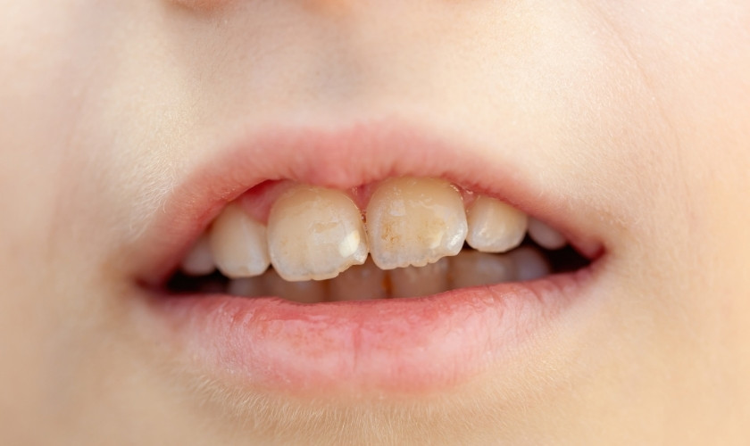
When a patient discovers opaque, milky patches on their teeth, the immediate concern is usually aesthetic. However, as a preventive care editor, I look at these marks as biological indicators of the tooth’s history and current environment. These dental white spots are not all created equal; they fall into three distinct categories based on when and how they formed.
The first, and perhaps most common for adults, is enamel demineralization. This is a post-eruptive process, meaning it happens after the tooth has entered the mouth. It occurs when the oral environment becomes too acidic, causing minerals like calcium and phosphate to leach out of the enamel. This often manifests as white spot lesions after braces treatment, because orthodontic brackets create tiny crevices where plaque can accumulate. If the plaque is not meticulously removed, the resulting bacterial acids create a porous surface that appears white and chalky.
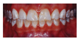
The second culprit is dental fluorosis. Unlike demineralization, fluorosis is a developmental condition. It occurs during the years when permanent teeth are still forming under the gums, typically from birth to age 8. According to data from the 2015-2016 National Health and Nutrition Examination Survey (NHANES), approximately 70% of U.S. children and adolescents exhibit some degree of dental fluorosis. This is caused by a systemic over-ingestion of fluoride, which interferes with the mineralizing cells and leads to increased enamel porosity.
Finally, there is enamel hypoplasia. This is a developmental disturbance that results in thin or irregular enamel. It can be caused by a wide range of factors, including nutritional deficiencies during infancy, high fevers, or localized trauma to a baby tooth that affects the developing permanent tooth underneath. This makes the affected teeth more prone to decay because the protective barrier is naturally weaker.
| Feature | Dental Fluorosis | Enamel Demineralization |
|---|---|---|
| Timing | During tooth development (pre-eruptive) | Any time after tooth eruption (post-eruptive) |
| Primary Cause | Excessive systemic fluoride intake | Plaque acid and poor oral hygiene |
| Appearance | Often symmetrical, lacy white streaks | Localized, often near gums or brackets |
| Reversibility | Structural change; requires surface treatment | Can often be reversed if caught early |
Diagnostic Checklist: Identify Your Spots
- Symmetry: Are the spots appearing in a similar pattern on both the left and right sides of your mouth? (Likely Fluorosis)
- Location: Are the spots located specifically where orthodontic brackets used to be or right along the gum line? (Likely Demineralization)
- Texture: Does the spot feel rough or "sticky" when touched by a dental tool? (Likely active Demineralization)
- Timing: Have the spots been there since the tooth first came in? (Likely Fluorosis or Hypoplasia)
The Science of Remineralization: Minerals and pH
Understanding the chemistry of your mouth is the first step toward long-term wellness. Our teeth are in a constant state of flux, shifting between demineralization (losing minerals) and remineralization (gaining minerals). The gatekeeper of this process is the oral pH level. Under normal conditions, saliva is rich in Salivary minerals like calcium and phosphate, which act as a natural repair kit for the enamel.
However, when we consume fermentable carbohydrates or sugary drinks, bacteria in the mouth produce acid. Once the oral pH level drops below the 5.5 threshold, the remineralization process stops, and the acid begins to dissolve the Hydroxyapatite crystals that make up our enamel. This is why reducing acidic foods to stop enamel demineralization is one of the most effective lifestyle changes you can make.
We can actively support this repair process by remineralizing teeth through diet. Focus on consuming mineral-rich foods like leafy greens, almonds, and dairy products, which provide the raw materials your saliva needs to protect your teeth. Additionally, staying hydrated with neutral-pH water ensures a steady flow of saliva to wash away acids and buffer the mouth back to a healthy state.
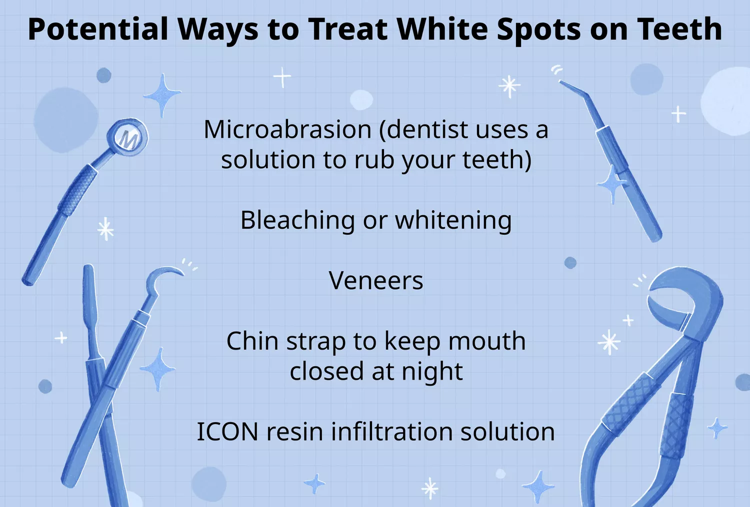
Professional & Home Treatments for Recovery
If you are wondering, can dental white spots be reversed, the answer is often a hopeful "yes," provided the lesions are caught in the early stages of demineralization. Once the surface of the enamel has actually broken into a cavity, remineralization is no longer possible, and a filling is required. However, for those "white scars" left after orthodontics or early-stage decay, several pathways exist.
For home care, your dentist might recommend calcium phosphate treatments for white spots. Products containing Casein phosphopeptide (CPP-ACP), often derived from milk protein, are highly effective at delivering bio-available calcium and phosphate directly into the porous parts of the enamel. Some patients may also be prescribed high-concentration toothpaste containing 5,000 ppm fluoride to jumpstart the remineralization process.
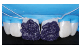
When home treatments aren't enough, professional clinical interventions offer impressive results. One of the most innovative methods is the Resin infiltration technique, often known by the brand name ICON. This treatment involves using a specialized resin that flows into the microscopic pores of the white spot. Because the resin has a similar light-refractive index to healthy enamel, it makes the white spot "disappear" by blending it into the surrounding tooth structure. This is a non-invasive, drill-free option that is particularly effective for white spot lesions after braces treatment.
For surface-level stains or mild fluorosis, a Microabrasion treatment may be used. This process involves a gentle mechanical removal of a very thin layer of the outer enamel using a mild acid and an abrasive paste. This can smooth out the "chalky" appearance and reveal the more translucent enamel underneath.
Prevention Strategies: From Infancy to Orthodontics
Preventing white spots requires a dual approach: managing systemic intake during childhood and maintaining local hygiene throughout adulthood.
For parents, dental fluorosis prevention is key. The prevalence of dental fluorosis among U.S. adolescents aged 12–15 significantly increased from 22.6% to 40.7% between the late 1980s and the early 2000s. To address this, the U.S. Public Health Service updated its recommendation in 2015, setting the optimal fluoride concentration in community water to 0.7 milligrams per liter. This level provides a protective benefit against cavities while minimizing the risk of spots.
When considering how to prevent dental fluorosis in children, parents should monitor toothpaste usage. For children under three, use only a "smear" (the size of a grain of rice), and for children ages three to six, use a pea-sized amount. Ensure they spit out the toothpaste rather than swallowing it.
For those currently wearing orthodontic brackets, the focus shifts to preventing enamel demineralization causes. Because brackets act as "food traps," standard brushing is often insufficient. Using an interdental brush or a water flosser is essential to clear away plaque from the edges of the brackets. Maintaining a high oral pH level by avoiding frequent snacking on acidic or sugary foods will keep the enamel hard and resistant to the "white scars" that often appear when braces are removed.
FAQ
What causes white spots to appear on teeth?
White spots are generally caused by one of two things: a loss of minerals in the enamel (demineralization) due to plaque acid, or an overexposure to fluoride while the teeth were still forming under the gums (fluorosis). They can also be caused by enamel hypoplasia, which is a developmental thinning of the enamel.
Can white spots on teeth be reversed?
If the spots are caused by early-stage demineralization, they can often be reversed or significantly improved through remineralization treatments like fluoride therapy, hydroxyapatite, or calcium phosphate. However, spots caused by fluorosis are structural and usually require professional cosmetic treatments like resin infiltration or microabrasion to change their appearance.
How do you treat white spots after braces?
The most effective professional treatment for white spots after braces is the resin infiltration technique, which fills the porous enamel and restores its natural look. Home treatments involving remineralizing pastes and improved hygiene can also help fade the spots over time if the damage is superficial.
Are white spots on teeth caused by too much fluoride?
Yes, when white spots appear as symmetrical, lacy streaks across many teeth and have been present since the teeth erupted, they are likely the result of dental fluorosis. This happens when a child ingests too much fluoride from water, toothpaste, or supplements before the age of eight.
Can white spots on teeth go away on their own?
Generally, white spots do not go away on their own without intervention. Demineralized spots may worsen into cavities if hygiene isn't improved. However, with a dedicated routine of remineralization and pH management, the enamel can harden, and the appearance of the spots can become less noticeable over time.
The journey to a brighter smile is as much about chemistry and biology as it is about hygiene. By understanding the difference between dental fluorosis and enamel demineralization, you can choose the right preventive steps. Whether it is adjusting your child’s fluoride intake or incorporating mineral-rich foods into your diet, these small daily routines build the foundation for long-term oral health and lasting confidence.