Quick Facts
- Survival Impact: Maintaining a regular schedule for a professional foot assessment can lead to a 36-86% reduction in amputation rates by catching issues before they escalate.
- Lifetime Risk: Current medical literature estimates that between 19% and 34% of people living with diabetes will develop a foot ulcer during their lifetime.
- The Golden Standard: A professional neuropathy foot assessment utilizing a 10g monofilament is the clinical benchmark for identifying a loss of protective sensation.
- Warning Sign: Any area of redness that spreads more than 2cm or is accompanied by warmth and swelling is a medical emergency requiring immediate clinical intervention.
- Amputation Catalyst: Statistics show that 85% of all non-traumatic lower-limb amputations in individuals with diabetes are preceded by a foot ulcer.
- Temperature Safety: Always check bath or foot-soak water with an elbow or thermometer to ensure it remains strictly below 37°C to prevent neuropathic burns.
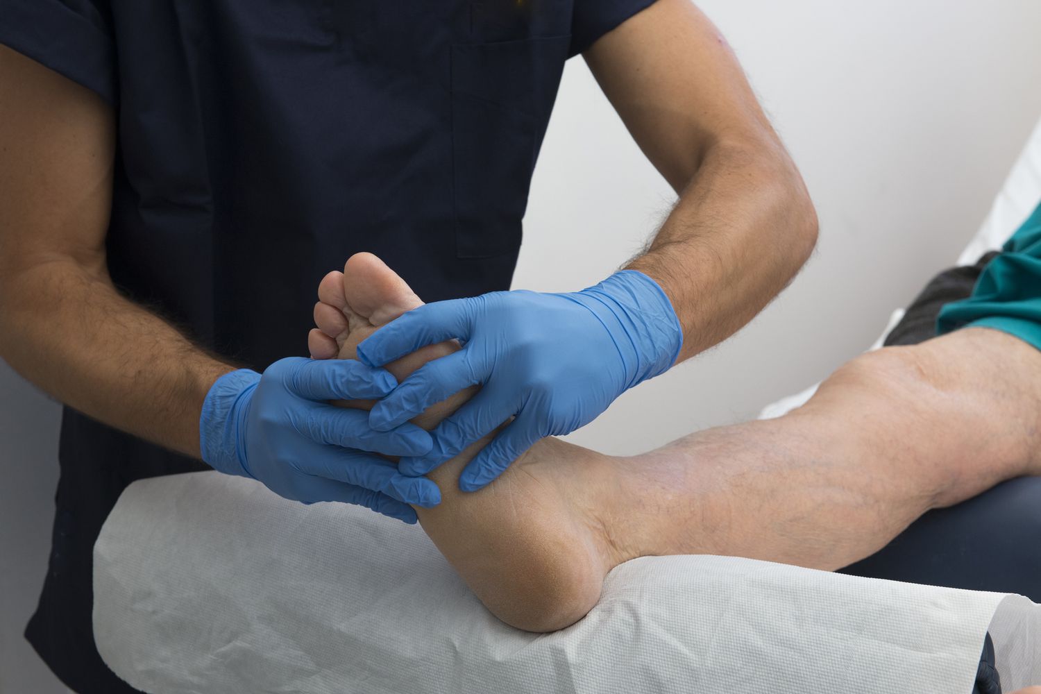
A professional diabetic foot exam is a comprehensive clinical assessment of skin integrity, vascular health, and neurological function used to identify risks for ulcers and infection. While daily self-checks are an important part of a wellness routine, the difference between diabetic foot screening and self-checks lies in the use of specialized diagnostic tools—such as monofilament testing and vascular studies—that detect invisible nerve damage and circulation issues before they become visible to the naked eye.
Why Self-Checks Aren't Enough: The Invisible Risks
As someone who focuses on preventive healthcare, I often tell my readers that the most dangerous complications are the ones you cannot see or feel. In the context of diabetes, the foot is a complex biomechanical structure that bears the brunt of systemic metabolic changes. While I always advocate for a nightly visual inspection using a mirror, relying solely on your own eyes can create a false sense of security.
The clinical triad of diabetic foot complications consists of neuropathy, peripheral artery disease (PAD), and infection. These three factors often work in silence. Peripheral Neuropathy, for instance, doesn't just cause a "pins and needles" sensation; it can progress to a complete loss of protective sensation. When this happens, a small pebble in your shoe or a blister from a new pair of loafers can go unnoticed for days, leading to a deep tissue infection.
Furthermore, autonomic neuropathy can affect the sweat glands in the feet, leading to excessively dry skin and cracking. These microscopic fissures in the skin integrity provide a gateway for bacteria. There is also motor neuropathy, which weakens the small muscles of the foot, leading to structural shifts like hammertoes or a collapsed arch, known as Charcot Foot. These internal biomechanical stress changes are often invisible to the patient until a callus or ulcer forms at a new pressure point. This is why preventing diabetic foot ulcers requires a professional eye that understands the underlying anatomy and can spot early warning signs of diabetic foot complications that the average person would simply overlook.
| Feature | Daily Self-Check | Professional Diabetic Foot Exam |
|---|---|---|
| Primary Goal | Spotting new cuts, blisters, or redness. | Detecting underlying nerve and vascular damage. |
| Tools Used | Mirror, good lighting, clean hands. | Monofilament, tuning fork, Doppler ultrasound. |
| Depth of Assessment | Superficial/Surface level. | Comprehensive (Vascular, Neuro, Musculoskeletal). |
| Detection Capability | Visible changes only. | Invisible ischemia and sensory loss. |
| Frequency | Every single night. | Every 1 to 12 months, based on risk level. |
The 5 Pillars: What to Expect During a Diabetic Foot Exam
When you step into a podiatrist's office or a specialized clinic for your screening, the process is much more than a quick glance. A high-quality diabetic foot exam follows five specific pillars of assessment to build a total picture of your lower-limb health.
1. Comprehensive Patient History The clinician will start by discussing your history of glycemic control and any previous instances of ulceration or foot surgery. They want to understand your daily activity levels and how your diabetes management has evolved over time.
2. Dermatological Assessment This is a meticulous check of the skin and nails. The clinician looks for fungal infections, thickened nails (onychomycosis), and calluses. In the diabetic foot, a callus is not just a patch of hard skin; it is a sign of repetitive biomechanical stress and is often a precursor to an ulcer.
3. Musculoskeletal Evaluation Here, the provider checks the structure of your foot. They are looking for deformities like bunions or hammertoes and assessing the range of motion in your ankle and toe joints. Limited joint mobility is a significant risk factor for increased plantar pressure.
4. Neurological Assessment This is where the monofilament test for diabetic foot screening comes into play. The clinician uses a small, flexible nylon fiber (a 10-gram monofilament) to touch various points on your foot while your eyes are closed. If you cannot feel the 10-gram pressure, you have lost protective sensation. They may also use a 128-Hz tuning fork for a vibration perception test to check the health of deeper nerve fibers.
5. Vascular Assessment Healthy blood flow is essential for wound healing. The provider will feel for your pedal pulses (the dorsalis pedis and posterior tibial pulses). If the pulses are weak, they may perform a vascular assessment for diabetic foot health using an Ankle-Brachial Index (ABI), which compares the blood pressure in your ankle to the blood pressure in your arm.

Risk Stratification: How Often Should You Be Screened?
The frequency of your visits shouldn't be a guessing game. Clinical diabetic foot screening guidelines provide a clear roadmap based on your individual risk category. By categorizing patients, healthcare providers can tailor the intensity of preventive care to those who need it most, effectively supporting the goal of limb salvage.
Red Flag Sidebar: When to Call Your Doctor Immediately If you notice any of the following between scheduled exams, do not wait:
- Redness that is spreading or exceeds 2cm in diameter.
- Drainage (pus or clear fluid) on your socks.
- A new foul odor coming from the foot.
- Systemic symptoms like fever or chills accompanying a foot sore.
- A sudden change in the shape or color of the foot.
The following schedule is generally recommended by wound care specialist professionals:
- Category 0 (Low Risk): No loss of protective sensation, no PAD, and no deformity.
- Frequency: Annual professional foot exam.
- Category 1 (Moderate Risk): Loss of protective sensation (LOPS) detected via monofilament, but no other complications.
- Frequency: Professional assessment every 3 to 6 months.
- Category 2 (High Risk): LOPS combined with either a foot deformity or signs of peripheral artery disease.
- Frequency: Clinical screening every 2 to 3 months.
- Category 3 (Highest Risk): A history of a previous foot ulcer, a lower-limb amputation, or end-stage renal disease.
- Frequency: Evaluation every 1 to 2 months.
How often should diabetics have a professional foot exam is a question that changes as your health status changes. If you develop a new callus or notice your shoes are fitting differently, you should move up your next appointment regardless of your current category.
Professional Footwear Evaluation and Home Care Integration
One often-overlooked aspect of a professional diabetic foot exam is the diabetic footwear evaluation during clinical exams. Your shoes are your first line of defense against the environment, but they can also be your greatest enemy if they don't fit correctly.
During a professional visit, bring the shoes you wear most often. The clinician will inspect the interior for protruding seams or worn-out cushioning. They may even suggest orthotics to redistribute pressure away from "hot spots." One holistic tip I love to share with my readers is the two-color pen shoe-tracing exercise. At home, stand on a piece of paper and have someone trace your bare foot with a blue pen. Then, place your favorite shoe over that tracing and trace the shoe with a red pen. If the blue line (your foot) is outside or even touching the red line (the shoe), that footwear is too narrow and is putting you at high risk for ulceration.
Integration of home care is the final piece of the puzzle. While a professional will handle debridement (the safe removal of calluses and dead tissue), you must resist the urge to perform "bathroom surgery" with scissors or razors at home. Instead, focus on the daily routine: washing with lukewarm water, drying thoroughly between the toes, and applying a high-quality urea-based moisturizer to the heels and soles (but never between the toes, as moisture there can lead to maceration and infection).
By combining the diagnostic power of clinical exams with diligent daily self-checks, you are taking the most effective steps toward long-term wellness and mobility.
FAQ
What happens during a diabetic foot exam?
A professional exam involves a multi-step check of your foot health. A clinician will review your medical history, inspect your skin for calluses or cracks, check the structure of your bones and joints, and test your circulation by feeling for pulses. They also perform neurological tests, such as the monofilament and vibration tests, to see if you have lost sensation in your feet.
How often should a person with diabetes have a foot exam?
At a minimum, every person with diabetes should have a professional foot exam at least once a year. However, if you have complications like nerve damage (neuropathy), poor circulation, or a history of foot ulcers, your doctor will likely recommend more frequent screenings, ranging from every month to every six months.
What are the warning signs of diabetic foot complications?
Look for early warning signs such as unusual swelling, persistent redness, skin that feels warm to the touch, or the appearance of calluses and corns. Other signs include changes in skin color, thickened or fungal toenails, and any open sore—no matter how small—that does not seem to be healing within a day or two.
Can I perform a diabetic foot exam at home?
You should perform a daily self-check at home to look for visible changes like cuts, blisters, or redness. However, a home check cannot replace a professional exam because you cannot accurately test your own nerve sensitivity or internal blood flow. Professional exams use specialized medical tools to identify risks that are not visible to the eye.
What is the 10-gram monofilament test for diabetes?
The 10-gram monofilament test is a simple, painless diagnostic tool used to check for a loss of protective sensation. A healthcare provider uses a small nylon fiber to apply a specific amount of pressure to different areas of your foot. If you cannot feel the fiber when it bends, it indicates that your nerves are damaged and you are at a higher risk for developing ulcers.
Why is a foot exam necessary for diabetic patients?
Diabetes can cause nerve damage and poor blood flow, making it difficult to feel injuries and slow for the body to heal them. Professional exams are necessary to detect these "invisible" problems early. Regular screenings are proven to reduce the risk of serious infections and can prevent the majority of diabetes-related amputations.